 3feeds.com
Dr. Flora Faure carefully places a small black cap on his head, resembling a swimming cap or the kind worn by rugby players.
The cap is dotted with hexagonal sensors that track brain activity.
At Rosie Maternity Hospital in Cambridge, researchers say they are the first in the world to test this technique, which could accelerate diagnosis and treatment for children with conditions such as cerebral palsy, epilepsy, and learning difficulties.
Experts suggest it might be available in UK hospitals within the next ten years.
“It’s the first time that light and ultrasound have been used together like this to give a more complete picture of the brain,” says Dr Faure, a researcher from the Fusion (Functional UltraSound integrated with Optical Imaging in Neonates) study.
Our brains undergo daily changes in the weeks before and after birth.
Brain injury in newborns is a leading cause of lifelong disability, and the NHS is currently implementing a program aimed at reducing such injuries during childbirth.
These injuries can impair the brain’s communication with the body, resulting in conditions like epilepsy, which causes seizures, or cerebral palsy, which affects movement and coordination.
While premature births increase the risk, brain injuries can also result from oxygen deprivation, hemorrhage, infection, or trauma during delivery.
For the roughly five out of every 1,000 babies affected, current monitoring techniques often struggle to predict the nature or severity of long-term impacts.
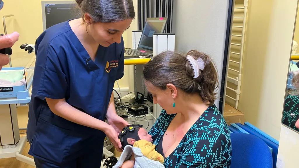
Explaining how the cap works, Dr Faure says: “The light sensors monitor changes in oxygen around the surface of the brain – a technique known as high-density diffuse optical tomography – and the functional ultrasound allows us to image the small blood vessels deep in the brain.”
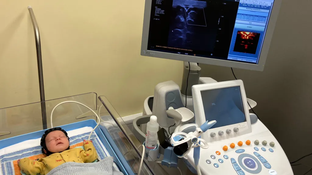
The device stands out because it is portable, allowing babies to be monitored more frequently and comfortably while in their cots.
Consultant neurosurgeon Dr. Alexis Joannides says it could offer several benefits compared with traditional MRI (magnetic resonance imaging) or cranial ultrasound (CUS) scans.
“MRI has limitations for two reasons: one is the cost and availability of scan slots,” he explains.
“The other is that you have to take the baby to a noisy scanner, wait maybe 20 minutes for the scan and then take the baby back again.
“It means, realistically, you can’t perform a series of scans, but in those first weeks, the brain can change daily so having a way of doing repeated tests is incredibly powerful.”
MRI and cranial ultrasound (CUS) are also seen as limited in predicting the type of impairment, due to the complex link between brain structure and function. However, a 2018 study by Imperial College London suggested that adding a brief 15-minute scan could improve their accuracy.
Regular testing of infants aims to identify issues much earlier, allowing therapies and interventions to start sooner.
The charity Action Cerebral Palsy has expressed support for the research.
“For many children with cerebral palsy, the road to diagnosis is a long one, and families can spend years knowing their child is ‘at risk’ of developmental issues but not fully understanding what that will mean,” says its founder Amanda Richardson.
“Technology like this could make all the difference, but it’s important that the capacity of community therapists is boosted to keep up with demand, as there is already a long wait for help.”
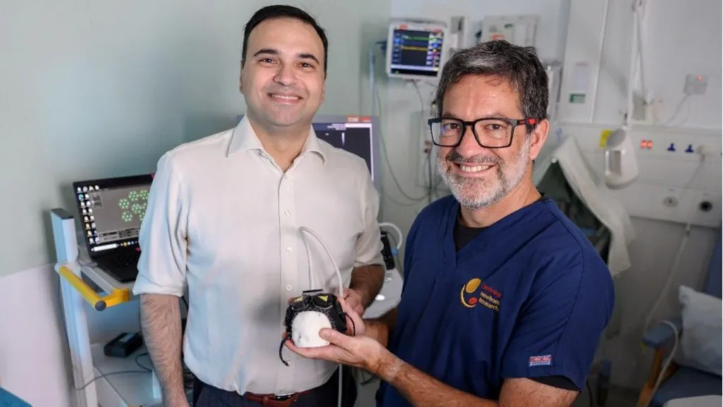
Professor Topun Austin, a consultant neonatologist, directs the Evelyn Perinatal Imaging Centre at Cambridge University Hospital. His research centers on brain treatments at both ends of life—infancy and old age.
He explains: “The Fusion study aims to develop and demonstrate a system for the cot-side assessment of brain activity in newborn infants and is currently the first of its kind in the world.
“We have spent 12 months successfully proving the concept with the help of healthy and premature babies and will now focus on babies considered to be at higher risk of brain damage.
“Understanding brain activity patterns in both term and preterm infants can help us identify those most vulnerable to injury at an early stage.”
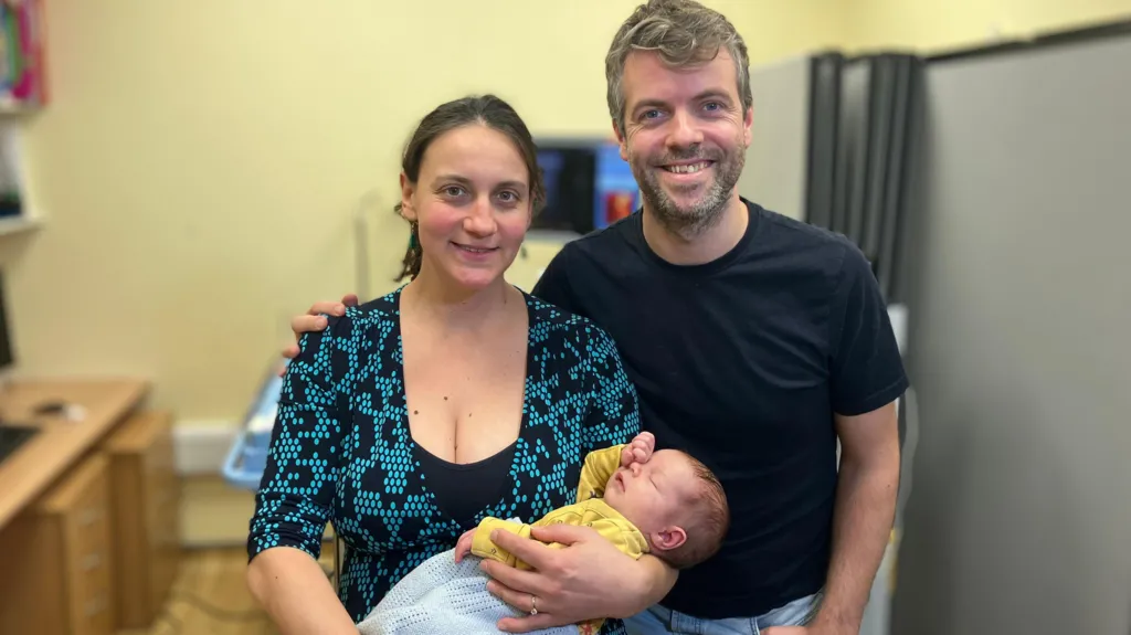
Theo is among the healthy full-term babies participating in the trial, and his mother, Stani Georgieva, believes it’s important to be part of the research.
“His dad and I are both scientists and when Theo grows up he’ll be able to take advantage of all of the advancements that have been made through research, so we felt it was important for him to be a little part of that understanding,” she says.
Dr. Joannides also serves as co-director of the NIHR HealthTech Research Centre in Brain Injury in Cambridge, which focuses on developing new technologies to enhance the lives of people with brain injuries.
The centre has funded a researcher for this study and will provide its expertise to help implement the device across the NHS if the trial proves successful.
“We still have hurdles to overcome, but we hope, within three to five years we’ll have a product that can be evaluated more widely,” he says.
“Cost permitting, it could not only monitor babies with a known problem, but also be a screening tool to help identify others who may be at risk.”
NIHR is leading a nationwide initiative to get more people involved in health research.